How to Treat Someone Who Complains of Chest Pains
How a patient who complains of chest pain is treated will depend on the diagnosis.
To make a diagnosis of chest pain, a health care provider will first get a detailed history of the exact location of the pain, where it radiates to, how long it has been on, how it started, any associated symptoms, any previous episodes and how it was resolved.
The clinician will also ask about history of high blood pressure, peripheral vascular disease, stroke, high cholesterol, high triglyceride, pneumonia, pulmonary embolism, pneumothorax, diabetes, kidney disease, stomach ulcer, reflux disease, gall bladder disease, liver disease, pancreatitis, chest injury, family history or heart attack, medication history especially aspirin and nitroglycerine use, etc.
Based on the outcome of this history, the clinician will conduct a general physical examination, followed by a focused chest examination.
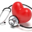
He/she will inspect, palpate, auscultate (with a stethoscope), for tender spots, swelling, heaves, fractures, abnormal heart sounds, abnormal breath sounds, abdominal tenderness, gall bladder tenderness, kidney and pancreatic tenderness etc.
The arms and legs will be examined for signs of stroke, while the eyes are examined for signs of internal bleeding in the head.
Next the patient will be hooked unto the ECG machine for an electrocardiographic recording to rule out ischemic heart disease or heart attack, which is the biggest threat.
Blood is also collected for cardiac enzyme and coagulation tests to further double-check for heart attack.
If there is a suggestion of heart attack, the patient is quickly admitted into the ICU.
Portable chest x-ray is done in ICU to rule out pulmonary embolism, pneumonia and pneumothorax.
A CT scan of the head and chest is also done to rule out stroke and pulmonary embolism.
Immediate anticoagulation treatments including aspirin are started if there is any suggestion of vascular obstruction in the heart or brain by a blood clot.
Nitroglycerin and selective beta blockers are initiated to increase blood supply to the hear muscles, if there is a high suspicion of heart attack.
Meanwhile the patient is put on oxygen mask and electronic heart and lung monitor.
The oxygen saturation, blood pressure, pulse, and respiratory rates are monitored for any negative changes.
Emergency resuscitation is kept handy for possible cardiac arrest or arrythmias (irregular hear beats).
The AED machine is kept handy in case the heart stops beating or starts beating irregularly or too fast.
By this time is would have become fairly clear where the chest pain is coming from.
If so the specific treatment is continued according to normal schedules.
Otherwise explorations of abdominal diagnostic options are commenced, to rule out gastric ulcer, PUD, esophageal reflux, all bladder disease and pancreatitis.
Detailed investigations are usually conducted when the patient is stabilized in ICU and transferred to the ward.
The rest of the treatment will then depend on subsequent findings and final diagnosis.
To make a diagnosis of chest pain, a health care provider will first get a detailed history of the exact location of the pain, where it radiates to, how long it has been on, how it started, any associated symptoms, any previous episodes and how it was resolved.
The clinician will also ask about history of high blood pressure, peripheral vascular disease, stroke, high cholesterol, high triglyceride, pneumonia, pulmonary embolism, pneumothorax, diabetes, kidney disease, stomach ulcer, reflux disease, gall bladder disease, liver disease, pancreatitis, chest injury, family history or heart attack, medication history especially aspirin and nitroglycerine use, etc.
Based on the outcome of this history, the clinician will conduct a general physical examination, followed by a focused chest examination.
He/she will inspect, palpate, auscultate (with a stethoscope), for tender spots, swelling, heaves, fractures, abnormal heart sounds, abnormal breath sounds, abdominal tenderness, gall bladder tenderness, kidney and pancreatic tenderness etc.
The arms and legs will be examined for signs of stroke, while the eyes are examined for signs of internal bleeding in the head.
Next the patient will be hooked unto the ECG machine for an electrocardiographic recording to rule out ischemic heart disease or heart attack, which is the biggest threat.
Blood is also collected for cardiac enzyme and coagulation tests to further double-check for heart attack.
If there is a suggestion of heart attack, the patient is quickly admitted into the ICU.
Portable chest x-ray is done in ICU to rule out pulmonary embolism, pneumonia and pneumothorax.
A CT scan of the head and chest is also done to rule out stroke and pulmonary embolism.
Immediate anticoagulation treatments including aspirin are started if there is any suggestion of vascular obstruction in the heart or brain by a blood clot.
Nitroglycerin and selective beta blockers are initiated to increase blood supply to the hear muscles, if there is a high suspicion of heart attack.
Meanwhile the patient is put on oxygen mask and electronic heart and lung monitor.
The oxygen saturation, blood pressure, pulse, and respiratory rates are monitored for any negative changes.
Emergency resuscitation is kept handy for possible cardiac arrest or arrythmias (irregular hear beats).
The AED machine is kept handy in case the heart stops beating or starts beating irregularly or too fast.
By this time is would have become fairly clear where the chest pain is coming from.
If so the specific treatment is continued according to normal schedules.
Otherwise explorations of abdominal diagnostic options are commenced, to rule out gastric ulcer, PUD, esophageal reflux, all bladder disease and pancreatitis.
Detailed investigations are usually conducted when the patient is stabilized in ICU and transferred to the ward.
The rest of the treatment will then depend on subsequent findings and final diagnosis.